
Written by: Eilidh McClain
Edited by: Olivia Pifer Alge, Mena Davidson, Kristen Loesel, and Jennifer Baker
Illustrated by: Jacquelyn Roberts
Start of experiment. Shift one. 3 hours in.
Cuckoo! “Scan finished.”
We hear the announcement that a scan is finished, sitting in a control room at Hamburg’s European X-ray Free Electron Laser. I wasn’t expecting it the first time; it’s such a silly little sound to use in a serious scientific lab. I began to associate it with success in the experiment we were conducting—another successful data collection scan under our belts. The sound of people typing diligently on computers and the gentle hum of discussion between the roughly ten scientists in the small room provides a rather calming backdrop to the experience. With each experiment, I am amazed at all that goes into setting up measurement scans. The control room computer screen is littered with endless computer screens, each monitoring important parameters of the experimental setup in the other room. The decisions are made by the visiting scientists, coming from multiple labs all over the world, but the equipment is monitored and handled by the three to four resident scientists at the facility.
Our world is limited – we are confined to the control room while a scan is taking place. The room where the experiment takes place is protected by a heavy door with a complex interlock system. Before the laser is let into the experimental room, a search takes place to ensure that nobody is left in the room before the door is closed. This is because being in the same room as the X-ray free electron laser can be deadly. I remember being trained in the search, walking quickly around the room and calling out in my loudest voice that the door was about to be closed.
We’ve started our 60-hour crunch, spending five 12-hour shifts underground collecting as much data as possible. It’s funny that we travel to many amazing places (such as California, Japan, or Germany) to access this equipment, yet we spend all of our time cramped up in a small room without any natural light!
Even though all of this time is spent underneath the surface of the Earth, we are excited and optimistic about our time here. We know we’ll get plenty of data, which can lead to important scientific discoveries. No matter where we find ourselves in the world, all of these scientists gathered here at this moment for one purpose and one purpose only: to use the big fancy laser.
Why is the ‘60-hour crunch’ necessary? It doesn’t sound pleasant to have back-to-back 12-hour days (or, sometimes, 24-hour shifts)!
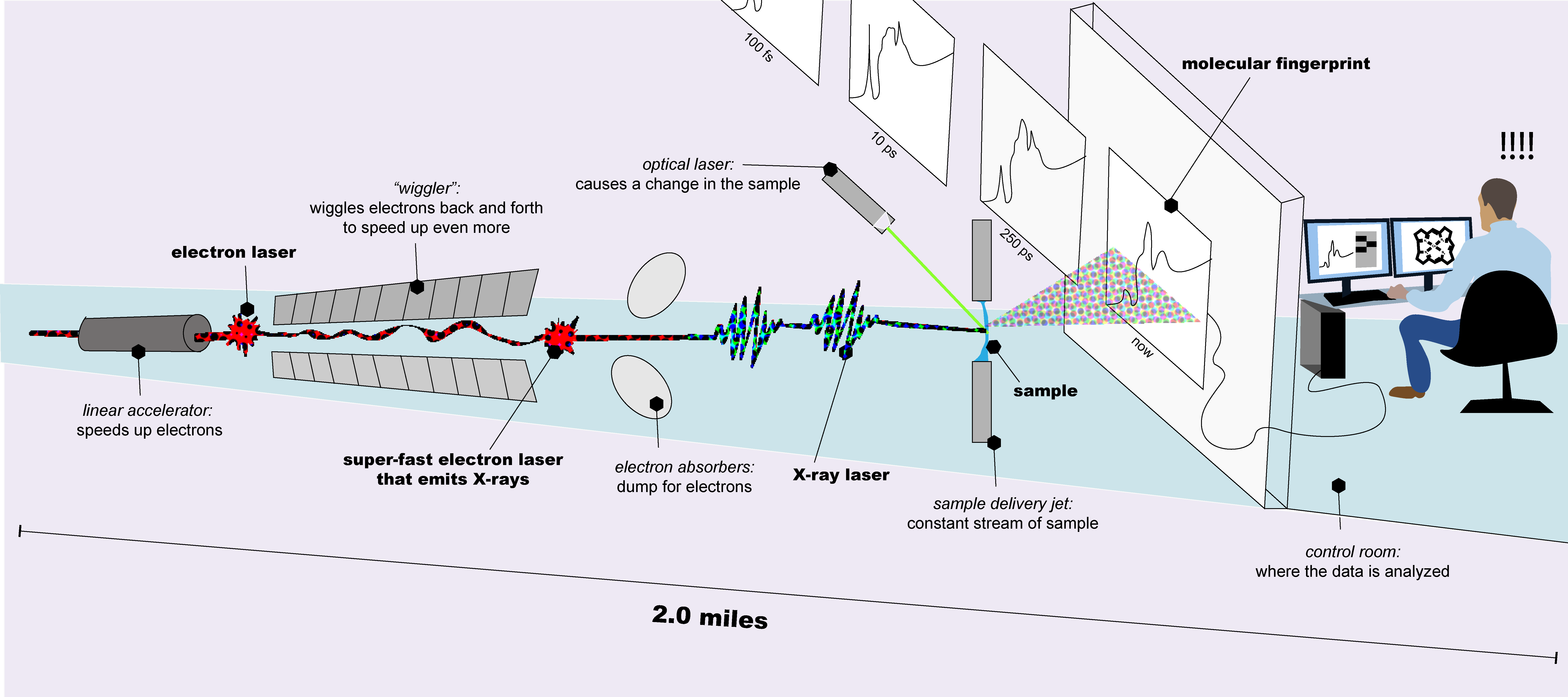
This ‘crunch’ is necessary because we are at an X-ray free electron laser (XFEL – pronounced ‘ex-fell’). An XFEL is a miles-long piece of equipment capable of experiments ranging from discovering the structure of proteins from the human body to simulating the extreme pressures and temperatures at the center of the Earth. In addition to being a powerful and versatile piece of equipment, the X-ray laser is made even more valuable by the fact that there are only five operating XFELs in the world! In order to do science with these facilities, one has to travel all over the globe. It’s a wonderful privilege to be able to do so, and since experimental time at an XFEL is a precious resource, we don’t want to waste even a few seconds of the X-rays if we can help it.
An XFEL works by accelerating electrons. At one end of the accelerator, an electron gun provides the electrons, which are flowed in a linear stream using strong magnets. Electronic motion releases energy according to the law of conservation of energy. This energy comes in the form of pulses, emitted in the X-ray region of the electromagnetic spectrum.
These pulses are very short, about 10-15 seconds long (one thousandth of one millionth of one millionth of a second!). Even though they’re very short, these pulses contain an incredible amount of energy. They are delivered in a fashion known as an ‘X-ray beam’. It’s like the beam emitted from a laser pointer, but invisible to our eyes and packing an extreme punch! The duration of our experiments is what we call ‘beamtime’, generally multiple days worth of time to obtain as much data as possible.
Shift two. Getting into a groove. 17 hours in.
Our experiment is underway. The first shift can be a bumpy ride, though the excitement of the beamtime helps to view first-shift problems through rose-colored glasses. By the second shift, we’ve become acclimated to the new state or country. In addition, the lab begins to feel more like home – we know our way around without too much guidance, and everyone’s picked their ‘assigned seat’ in the control room. It’s a silly little thing, but I find it comforting in a lab so far away from home. This comfortable feeling will only grow over our time here.
After having worked through most of the experimental kinks during the first shift, the second shift often feels like the official beginning of our data collection. The next challenge is to condense the huge amount of information from the experimental detectors and equipment into pieces that we can easily review. Analyzing data during the experiment allows us to make informed decisions, so as not to waste any beamtime. A time crunch like this is a computer programming-heavy adventure that gets us ever closer to the discoveries we yearn to make with this giant pulsed laser.
Pulsed lasers have common applications in everyday life, such as tattoo removal and Lasik eye surgery, among other uses. In the case of the pulsed X-ray laser, the discoveries are endless – it would be very difficult to enumerate them all here, because the applications of the XFEL are quite interdisciplinary. The discoveries range with everything from studying the structure of proteins to seeing how materials respond to extreme stress. Along with scientists from many different fields in the natural sciences, there are engineers, data scientists, and technicians who all work together to make a beamtime a success. Without the amazing scientists at the XFELs, many experiments would be impossible to conduct. The focus of my experiments is biophysics, a field that studies biomolecular structure and light-dependent reactions, in addition to other physical or chemical applications. However, it is very common for multiple types of samples and study goals to be addressed during a single beamtime.
Structural determination of biomolecules
In the world of biophysics, understanding the structure of biomolecules like proteins is very important. You may be familiar with the ‘central dogma of molecular biology’: replication of DNA, transcription of RNA, and translation into protein. In biophysics there is also a ‘central dogma’ of sorts. This is: structure determines function. In other words, the way that proteins and RNAs are folded in cells directly links back to the overall function of those biomolecules. This is why many researchers in biophysics are concerned with structural determination. The information we can get from the way a protein is folded can explain how that protein is working in the body – what does it do, and how does it do that?
XFELs have made this process faster and have allowed scientists to study how the structure of a protein changes over time within a given cellular reaction. One example of this is time-resolved serial crystallography. Crystallography is based upon the principle of X-ray diffraction, the same technique that helped Rosalind Franklin discover the alpha-helix structure of DNA. It’s like taking an ‘X-ray picture’ of a molecule. X-rays strike biomolecular crystals and diffract, forming circular dotted patterns that can then be converted into atomic structures. Time-resolved serial crystallography combines the power of the XFEL and these crystallographic techniques. Because the pulses from the XFEL are so short and so intense, there isn’t any measurable X-ray damage to the sample, unlike with other equipment. This principle is called ‘probe before destroy’. While the sample is eventually destroyed, the probing is so fast that this destruction is not detected, creating a valuable window into the characteristics of these samples.
Time-resolved serial crystallography takes snapshots of crystallographic data at different time points during a reaction to see how the structure of the protein changes as the reaction takes place. This data can then be used to create a ‘macromolecular movie’ of sorts, where a researcher can visualize parts of the protein changing position or folding structures emerge and disappear. Overall, this is important because half the battle of understanding how proteins and other biomolecules work in the body is understanding how they are folded and how they interact with other molecules around them. This information can lead to new drug targets, treatment options, and more.
Molecular movies
Speaking of movies, researchers can also take videos of much smaller molecules than proteins using an XFEL. In particular, time-resolved X-ray absorption spectroscopy (a measure of how much X-ray is absorbed by a molecule over time) can reveal the structure of a molecule. For example, in this paper, X-ray absorption spectroscopy is used to sequentially observe structural changes of the vitamin B12 analogue methylcobalamin. Methylcobalamin is one of the forms of vitamin B12 used in the body – it helps in the synthesis of amino acids necessary for building proteins in the body. Without it, the body can experience buildup of substances related to this process, a condition called homocystinuria. In a time-resolved X-ray absorption spectroscopy experiment, the methylcobalamin molecule starts with a certain structure. After perturbation with a light pulse, we can observe that molecular structure changing and distorting, allowing us to better understand the molecule’s reaction to light with a structural ‘molecular movie’.
This movie is important because light-mediated reactions are potentially non-invasive methods of drug delivery and reaction control. Imagine going to receive a treatment for a condition and rather than having to take multiple doses of a drug, the dosages are instead controlled by exposure to light in repeated cycles. In addition, light can allow for very site-specific treatments in cases where localized treatment is ideal, like treating cancerous tumor cells. Molecules like methylcobalamin are well-suited for this application as it is relatively easy to tack on different ligands to the molecule. Understanding how this molecule responds to light is paramount for attempting to tune this response in our favor.
Matter in extreme conditions
Now we venture away from biology and chemistry into the world of extreme physics. XFELs also allow researchers to study high-energy conditions that would be impossible to replicate on a large scale (unless you shot something into the sun, perhaps). In one case, researchers at the Matter In Extreme Conditions experimental station at California’s Linac Coherent Light Source (LCLS) discovered that the high pressures and temperatures in the center of ice giants like Neptune could create the fabled ‘diamond rain’ with the types of materials found on the planet. Using the hydrocarbon plastic polystyrene (C8H8), as both carbon and hydrogen are abundant on planets like Uranus and Neptune, researchers simulated the conditions deep inside the planets by inducing high temperatures and pressures with a powerful optical laser. They then probed what happened to the plastic with the X-ray pulses from the LCLS as the extreme conditions separated the carbons and hydrogens in the plastic from one another. They observed diamonds forming! This is just one example of the capabilities of this instrument at the LCLS. Research like this can help us better understand our solar system and our own planet.
Shifts 3 and 4. A few road bumps here and there, reaching the home stretch. 38 hours in.
We are in the thick of the experiment now. We are halfway through the beamtime – and, if we’re lucky, we’ll have more data than we know what to do with. But even now issues can arise, making these experiments very difficult and making every second precious. Because of the sensitivity and complexity of this instrument, it is not uncommon for a research team to experience one or more ‘beam dumps’ per beamtime. A beam dump occurs when one or more of the components in the miles-long laser experiences an issue and X-rays stop being delivered to the experimental area. I’ve experienced a few beam dumps on each experiment I’ve participated in. One in particular affected my data. It was the last day of the beamtime and the beam dropped after several hours of the shift, during a scan of one of my samples. There’s nothing to do in those circumstances except rely on the engineers and scientists who are qualified to fix the broken components, waiting anxiously for the beam to return. In this particular case, the beam didn’t come back for the rest of the shift, and that’s where our data collection ended, preventing me from collecting data on my sample. It is a bit disappointing when this occurs, and some groups lose whole shifts because of laser problems, but that is part of the reality of working with such a complex instrument.
The beam dumps can be addressed with future XFEL advancements, however. The speed of data collection is limited by the repetition rate of the pulses – in other words, how many pulses can be generated per second. This means that when a beam dump occurs, it’s not nearly as stressful, since the same amount of time can stretch much further. A successful, complete experiment requires about four to six 12-hour shifts at LCLS. Sometimes shifts are 24 hours and, while it’s difficult to monitor an experiment for so long, the longer shift reduces setup time. There’s a tradeoff between comfort and efficiency, for sure. It takes time to get lasers lined up, set up the experiment, and put the sample of interest into the beam. Therefore, being able to generate more pulses per second means researchers can acquire the same amount of data very quickly.
The current LCLS operates at 120 Hz, generating 120 pulses per second. The current fastest repetition rate is at Hamburg’s European XFEL with 27,000 pulses per second; however, they are normally delivered in ‘bunches’ at lower repetition rates. This means that more X-rays are delivered to the sample, but the speed of data collection is similar to that of the LCLS and other XFELs like it, with the same disadvantages. However, there is a solution on the horizon, with two new XFELs slated to be commissioned in the coming years. One is in China: SHINE, which is predicted to come online in 2025. The other is in California, USA: LCLS-II. LCLS-II is being commissioned as we speak, with “first light” expected in September 2023 and the first full experiments beginning in January 2024. The ramping process to full operational status will take a couple of years. But think of all the data that can be collected when it’s complete!
At its full potential, the new LCLS-II system will operate at 1 MHz, with a million pulses per second. Imagine the reduction of experimental time – more experiments can be performed faster and more efficiently. Beamtimes likely won’t last nearly as long, avoiding the dreaded, caffeine-laden 24-hour shifts. Researchers will likely be less grumpy by the end of it all and be incredibly happy with the quality of their data!
Shift 5. The last day. 59 hours and 58 minutes in.
We’re exhausted but also looking forward to leaving the underground experimental stronghold and walking out into the light with new data and discoveries on the horizon. It’s not uncommon for research teams to celebrate the end of a shift with a celebratory meal or a collection of everyone’s favorite snacks. No matter what happens during the beamtime, we can only be satisfied with the new successes and breakthroughs we witnessed and helped create.
And, with that, our 60 hours are nearly up. From our rose-tinted first shift, through the struggles of the middle, and aiming towards our greatest chance of success, we can finally breathe at the end of our last shift. We will celebrate, and then all go our separate ways. Our groups will meet again later to talk about processing and analyzing the data we collected, and how it impacts the scientific question at hand.
End of beamtime. Congratulations!
XFELs are powerful instruments pushing the boundaries of many different scientific disciplines and aiding in new discoveries. The time is precious, but worth it, as even one trip could potentially generate the data needed for a PhD student to write their thesis! Current XFELs are valuable instruments, but they have limitations that are largely addressed by the generation of higher pulse repetition rates. So, whether you want to watch a molecule dance, determine how a protein is arranged, or study what it’s like at the center of the Earth, XFELs will continue to make this possible.

Eilidh McClain is a PhD candidate in the Biophysics program, studying vitamin B12 using lasers of various types, including that of XFELs like the LCLS. Eilidh graduated with an undergraduate degree in physics in 2019 from Kansas Wesleyan University. In her free time, she writes voraciously (mostly novels and short stories), learns languages, plays with cats, walks around, and collects tiny treasures. She believes the art of science communication is very important and is considering a career in this field once she graduates with her PhD. Additionally, she is grateful for her liberal arts education in giving her a good writing and presenting foundation with which to spread the magic of science to others.
