
Written by: Kate Giffin
Edited by: Elise Tahti
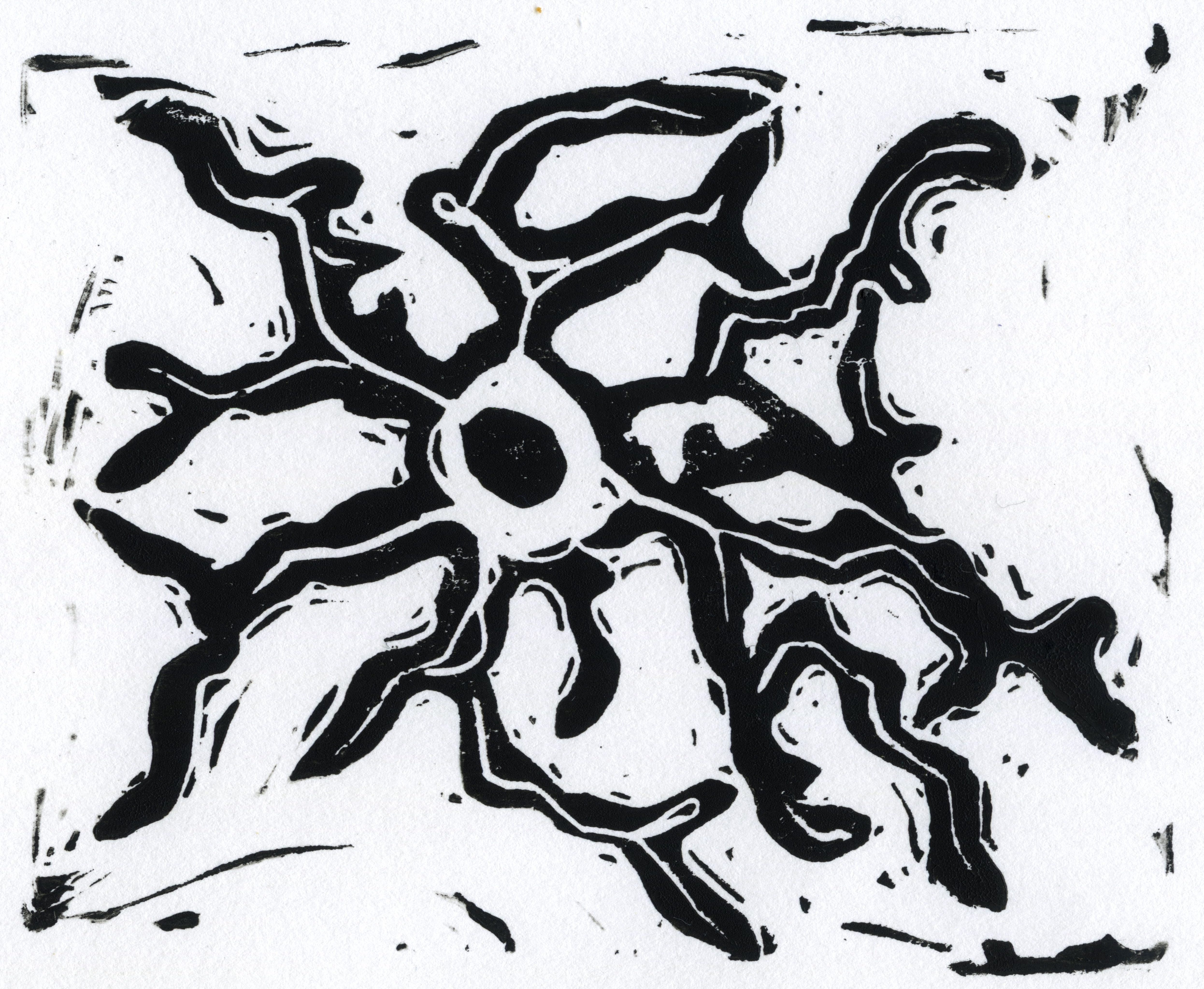
The first neuroscientists were artists. Using clever chemistry, they developed ways to stain brain tissue and then drew what they saw. Here, I have carved a block print based on one of the first published drawings of microglia, the immune cells of the brain. In a series of four 1919 papers, Pío del Río-Hortega described these cells and clearly showed that they were a different cell type from the other glial cells in the brain, astrocytes and oligodendrocytes. His conclusions about microglia, based almost solely on observations about how they look under different conditions, were startlingly accurate. For example, he noticed that microglia rapidly migrate to a site of injury and transform in shape, even becoming, in his words, “monstrous.” He accurately hypothesized that they clean up debris through a process called phagocytosis. He also speculated that microglia have a different developmental origin than other brain cell types, a conclusion that wouldn’t be verified for over 100 years and which required the development of sophisticated genetic fate mapping techniques.
Current neuroscientists still visualize microglia in health and disease to answer important scientific questions. But instead of pencil and paper, we use high-powered microscopes and lasers.
These are microglia from mouse brains in a model of multiple sclerosis, used to study the brain inflammation that contributes vision loss in multiple sclerosis. The colors represent proteins for normal microglial function (green; Tmem119) and disease-associated microglia (pink; Clec7a). Image credit: Jenna Staples from the lab of Sebastian Werneburg.
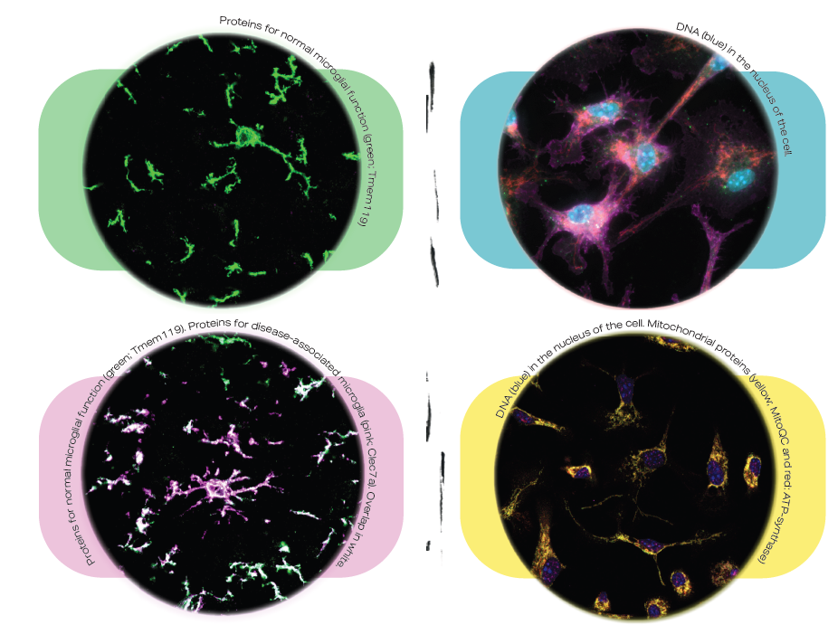
These are microglia that are grown in a dish and used to study cellular damage during brain injuries like stroke. The colors are for the DNA in the nucleus of the cell (blue/purple; DAPI) and mitochondrial proteins (yellow; MitoQC and red; ATP-synthase).
Across hundreds of years, microglia inspire art and scientific discovery. While the tools may have changed, the beauty of biology remains.

Among other things, Kate Giffin is a neuroscientist who recently defended her PhD. In her PhD, she studied how severe infections can lead to long-term brain issues like dementia, with a focus on the immune cells of the brain, microglia. She is passionate about telling scientific stories through unexpected genres to expand the way people think about science and the world. Recently, she has been learning new artistic mediums like linocut, watercolor, and zines. When Kate is not marveling at the brain, she is probably outside marveling at some strange plant.


