
Author: Ellen KW Brennan
Editors: Emily Glass, Sophie Hill, and Lisa Pinatti
Illustrator: Katherine Bonefas
Neurons are the main communicators of the brain. Using electrical signals to ‘talk,’ their conversations with each other underlie every behavior, thought, and feeling we have. To produce these larger functions, neurons need to work together in networks. For example, there is a specialized network of neurons whose only job is to keep you oriented in your surroundings. Two of the main types of neurons in this network are ‘place cells,’ which tell your brain where your body is in space, and ‘head direction cells,’ which tell your brain which way your head is facing. Together (and with the help of many other cells), they act as your body’s GPS system. While knowing where you are is important, you also need to know the details of the world around you. Other specialized cells, like sensory neurons called ‘blobs,’ help you detect color, while neurons in your nose called ‘olfactory sensory neurons’ catch chemicals as you breathe to detect smells. Together, these and many other different neural networks give us a sensory representation of our surroundings.
Another important reason to stay in tune with our neurons is that they can be indicators of many diseases. For example, one of the first detectable signs of Alzheimer’s Disease is ‘hypometabolism’ in parts of the cortex and memory areas of the brain. This means that these areas are less active—in other words, the neurons there have changed their communication within the network. These changes can serve as red flags that alert patients and doctors to begin treatments early in the disease course. For many neurodegenerative diseases with no cure, such as Alzheimer’s disease, early detection and treatment can add years of health to someone’s life.
But how did scientists discover that some neurons are specialized to tell us where we are in space while the communication patterns of others can foreshadow Alzheimer’s?
They listened to them.
Scientists eavesdrop on the conversations of these neurons to learn exactly what they say to other neurons and when. However, this can be a daunting task, as neurons are so small that they’re measured in microns, or 1/1000th of a millimeter, and it is difficult to attach a recording device to something this small. So how can we tap into the conversations of these tiny neurons?
Techniques such as functional MRI allow researchers to see what regions of the brain are increasing in activity or going quiet, while EEG lets them view the larger waves of signals flowing across the brain. Neither technique, however, can tell scientists exactly which cells are talking or what they’re saying. The first technique to achieve that level of detail is the patch-clamp, which involves infiltrating a tiny neuron with an electrode to record its electrical signals without harming the cell itself. While that may sound pretty tricky, this technique had humble beginnings over two centuries ago in the leg of a frog.
In 1797, physicist Luigi Galvani was nicknamed the “frogs’ dancing master” because he discovered that electrical signals from neurons caused the leg muscles of the frog to kick. By connecting the neurons in the frog spinal cord to the muscle of its disconnected leg using simple wires, he was able to make the leg contract. Galvani was the first to show that one neuron could send an electrical signal to a different neuron that could then send its own signal to the muscle and tell it what to do. Essentially, he discovered that neurons talk to each other and communicate via electrical excitation. He claimed that positive and negative charges inside and outside of the neuron create a flow of electrical current that produces the electrical signals neurons use to communicate. Turns out, Galvani was right, but what does that have to do with eavesdropping on neurons?

Well, that electrical current is exactly what neuroscientists use to know what neurons are saying to each other, but it took them a long time to figure that out. Galvani’s findings on “animal electricity” dominated this field of single-cell electrophysiology for over a century until zoologist John Young decided to take it to a new animal – the North Atlantic squid. In the 1930s, Young discovered that the movement of the squid depended on the giant axon, which is so large you can see it with the naked eye. Young realized that electrical signals from that giant axon told the squid’s body when to propel forwards and glide through the water, but he didn’t recognize its potential for studying how neurons talk to one another. Instead, that discovery would belong to two physiologists and neuroscientists, Alan Hodgkin and Andrew Huxley, who were investigating the squid axon as well. However, their work was delayed after their laboratory and research was nearly destroyed during World War II. It wasn’t until 1952 that they discovered the electrical basis of neuronal communication, termed the ‘action potential.’
Hodgkin and Huxley were one of the first research teams to make an electrode that could be inserted into the squid’s giant axon to record its electrical properties. Made of two silver wires, this electrode recorded the electrical signal that the neuron used to talk to other neurons in its network. Using that information, Hodgkin and Huxley concluded that an action potential is caused by a flash flood of positively-charged sodium ions entering the neuron at once, causing that positive charge to shoot down the neuron’s axon to the next neuron in their game of telephone. Their discovery of the fundamentals of neuronal communication was so critical to neuroscience that they were awarded the Nobel Prize in 1963.
For the next twenty years, scientists continued wiretapping into neuronal conversations by sticking them with wires to record their electrical signals. One key innovation was the sharp electrode, a glass spear used to penetrate the neuron and protect the recording wire that sits inside. While the wire was safe, the now-impaled neuron had a few issues. Over a relatively short amount of time, the neuron would die as its ions and nutrients leaked out of its open wound until the information being recorded was no longer accurate. This made recordings using sharp electrodes reliable but unstable, as stabbing a neuron tends to have detrimental effects on its health.

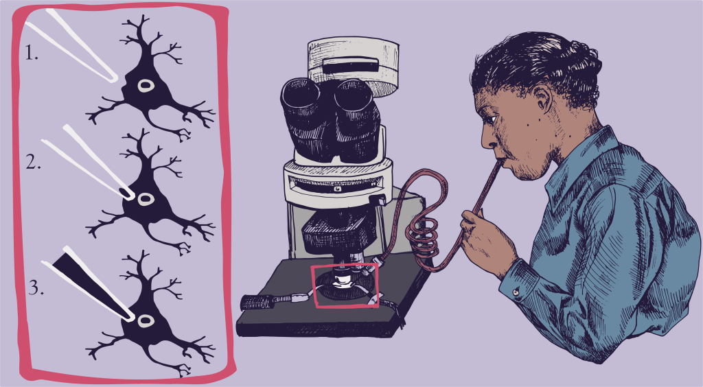
In order to solve this issue, biophysicist Erwin Neher and cell physiologist Bert Sakmann invented the patch clamp technique in the early 1980s, which enables scientists to record neurons in a more controlled manner. Instead of impaling the neuron with a sharp spear, a glass pipette with a wider opening is used to slowly approach the neuron until the pipette pushes against the neuron’s edge, very gently, to form a dimple. Then, by applying a small amount of suction, the outside wall of the neuron is drawn into the glass pipette until it forms an extremely strong seal. Picture a scientist using a long straw to gently slurp up part of the neuron – that’s exactly what they do.
After the seal is formed, the pipette is still outside the neuron. In order to get it inside where the electrical charge can be recorded with extreme detail, a sudden, strong amount of suction is used to break the part of the neuron’s outer wall that was sucked up into the pipette. Many electrophysiologists call this the “kiss of life,” partly because that suction noise sounds like a kiss and partly because they actually use their mouths to do it. Now the edge of the glass pipette remains attached to the surrounding intact wall of the neuron, keeping the neuron’s insides inside where they belong, while within the pipette, the electrode wire has become a continuous part of the neuron. Finally, scientists can record everything inside the neuron without causing a gaping wound, allowing them to gather accurate and informative data on how individual neurons communicate within their network.
Though it took one and a half centuries to develop, this same patch-clamp technique is still what researchers use today to approach many different questions about how the brain works. In this age of rapidly developing technology, there are still scientists sitting in lab slurping up neurons with their straws to get a quick listen into the inner workings of the brain. Using this humble technique, they’ve been able to discover new types of neurons, map the connections of the brain, identify early predictors of Alzheimer’s disease, develop new electrical treatments for diseases like Parkinson’s disease, and so much more.

Ellen KW Brennan is a PhD candidate in the Neuroscience Graduate Program at the University of Michigan. As an NSF GRFP scholar, she studies the electrophysiological properties and connections of cortical circuits involved in memory. Outside of the lab, she is passionate about both science communication and breaking down stigmas about mental health. Her dream is to work in science communication, bridging the gap between universities doing cutting-edge research and the public who funds them. When she isn’t focusing on science, you’ll find her performing improv, playing with her dogs, or cooking up something new.
